Diagnostics
Investigation of your complaints
Getting to the bottom of health problems
Orthopaedic diagnostics deals with various measures for the detection of different diseases. As the causes of diseases vary greatly, the diagnostic procedures vary considerably – depending on the complexity of the health problem and the suitability of the methods for the patient.
Typical procedures include anamnesis, clinical examination, sonography (ultrasound) and detailed examinations using imaging techniques such as MRI, CT and X-ray. We will discuss the procedures suitable for alleviating your condition in detail in a personal consultation at our private orthopaedic practice “ORTHO-EINS” in the Berlin district of Dahlem. We explain both positive and negative aspects of the state-of-the-art technical procedures as well as the orthopaedic approach.

Overview of diagnostic procedures
Sonography (ultrasound)
Ultrasound imaging (sonography) is often used to examine joints, muscles and tendons. For example, we examine the function of a muscle during contraction or relaxation and the joints during flexion or extension. In addition, the identical body part (e.g. opposite shoulder) can easily be used as a reference object. The results are included in the findings.
When are ultrasound examinations carried out
Ultrasound examinations are carried out in particular for shoulder and knee joint complaints, pain in the Achilles tendon, after muscle injuries and to check the progress of injuries (including joint and bruising). This painless examination is inexpensive and takes very little time. This does not result in any radiation exposure.
Dr. Topar also performs precise injections under ultrasound guidance.
X-ray
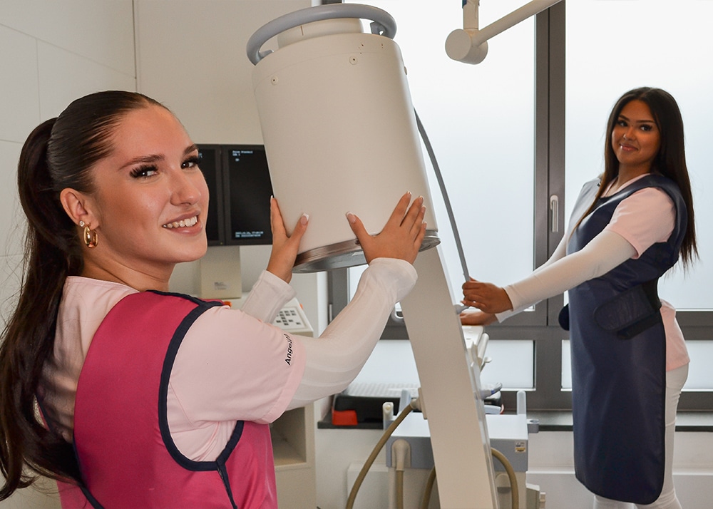
X-rays are a tried-and-tested and frequently used tool for detecting physical abnormalities, injuries and degenerative diseases. Our state-of-the-art device is at the cutting edge of science and therefore emits less radiation. Our expert staff operate the appliance in accordance with the specified guidelines for every job. The diagnosis can therefore be made without further delay.
Magnetic resonance imaging (MRI)
As back specialists, we use the imaging procedure of magnetic resonance imaging in our practice. This method is based on magnetic fields and alternating electromagnetic fields and does not use ionizing radiation (e.g. X-rays). This examination method is therefore radiation-free. MRI can be used to produce cross-sectional images that enable precise imaging of the human body and organs. Particularly soft tissue (such as intervertebral discs, meniscus, muscles) or bony structures can be displayed layer by layer in images on a monitor. We then make a precise diagnosis.
With the help of a state-of-the-art in-house magnetic resonance tomograph, it is also possible to take images on the very first day of your visit to the practice and initiate appropriate treatment – promptly and thoroughly.
Laboratory / blood test
To rule out or confirm a specific diagnosis, blood tests often play an important role in orthopaedics diagnostics. The determination of blood values is particularly important for rheumatic diseases. Blood tests are also important for the diagnosis and monitoring of other inflammatory or infectious diseases. In addition to blood, punctates (e.g. joint effusions) can also be diagnostically informative.
Corresponding samples are taken in our practice and analyzed in a central laboratory as quickly as possible.


